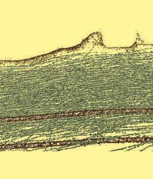
A Look into the Smallest Things
Cryo-electron microscopy
MikroMakro Parcours
Imagine you could see what is going on within our complex organism, from cellular tissue, over single cells, to their tiniest components like organelles, and even beyond that: individual proteins and biomolecules.
One efficient method for understanding how small things are integrated in bigger ones has revolutionized structural biology in the past few years: cryo-electron microscopy. Its name derives from the ancient Greek words κρύος (kríos, for cold), μικρός (mikrós, for small), and Σκοπιά (skopia, too look or look-out). The development of this technique over the last decades was honored with the 2017 Nobel Prize in chemistry.
A vitrified biological sample (of macro-, micro-, or nanometer size) is subjected to a high-energy electron-beam (300 kV) at -190 °C and high vacuum. The electron beam passes through the sample and a series of lenses and filters and finally hits a camera. Thereby, the electron beam penetrates the specimen and thus it is possible to visualize the interior layers of a sample, and not only its surface. In modern microscopes, the sample can furthermore be rotated resulting in cryo-electron tomography.
Cryo-electron tomography allows scientists to visualize and display 3-dimensional structures of biomolecules in high resolution. This knowledge of the structure and organization of proteins facilitates the development of drugs used to treat a variety of illnesses, from cancer to Alzheimer’s disease.
Bruno Martins is a PhD-student in the Research Group of Prof. Ohad Medalia at the Biochemical Institute at the University of Zurich. More
Related contributions
