A Voyage into the Cell

At the Department of Molecular Mechanisms of Disease (DMMD), University of Zurich, we combine cellular and molecular biology with biochemistry to better understand the way different cellular functions affect the development of diseases and their therapy.
Cellular and molecular biologists at the DMMD work together to develop various ways of illuminating and understanding the structures and processes that exist inside cells. We work hard to better understand the cell, the smallest subunit of our bodies. If something goes wrong in our cells, the whole body suffers. Numerous diseases are the result of the molecular processes inside our cells somehow being disturbed. Malfunctions that occur at the level of our DNA, our genes, and their regulation can have particularly serious consequences. For example, the cellular metabolism can be disturbed to such an extent so that it develops into diabetes. Mutations in the genetic material can make it easier for cancer to develop. A deregulated response on the part of the immune cells can lead to chronic inflammation. And if gene regulation isn't working properly, some cells may not even remember what their job is inside the body. Because cells are so small (one cell is about 100 times smaller than a pinhead), biologists need microscopes in order to look at them. Or they use other, sometimes quite complicated, imaging procedures to look inside the cells and watch the molecular mechanisms at work. These procedures are carried out in a research laboratory, they often take several days, and they're based on a very precise sequence involving numerous steps. If the experiment works, we know a little more about the cell. Based on the new findings, we can then make plans for subsequent experiments. We've yet to run out of new questions.
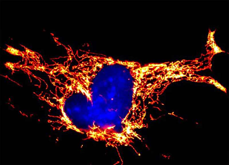 Through what's called immunostaining, the cell nucleus and mitochondria, which are the cellular powerhouses, can be rendered visible. Image taken by Ann-Katrin Hopp, research group Hottiger.
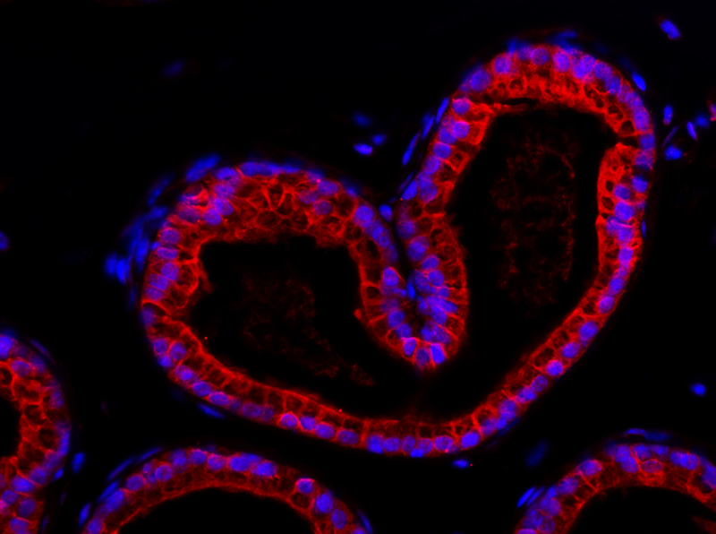 Cellular assemblies are the basis for organ formation. Sometimes interesting shapes are produced, such as this heart-shaped collection of prostate cells.
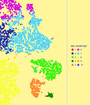
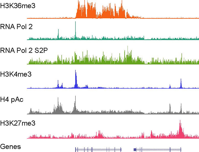 Genome-wide measurements of epigenetic modifications and computer-assisted analyses allow us to better understand how, specifically, genes are switched on and off. Measurements and analysis by Christina Ambrosi, research group Baubec.
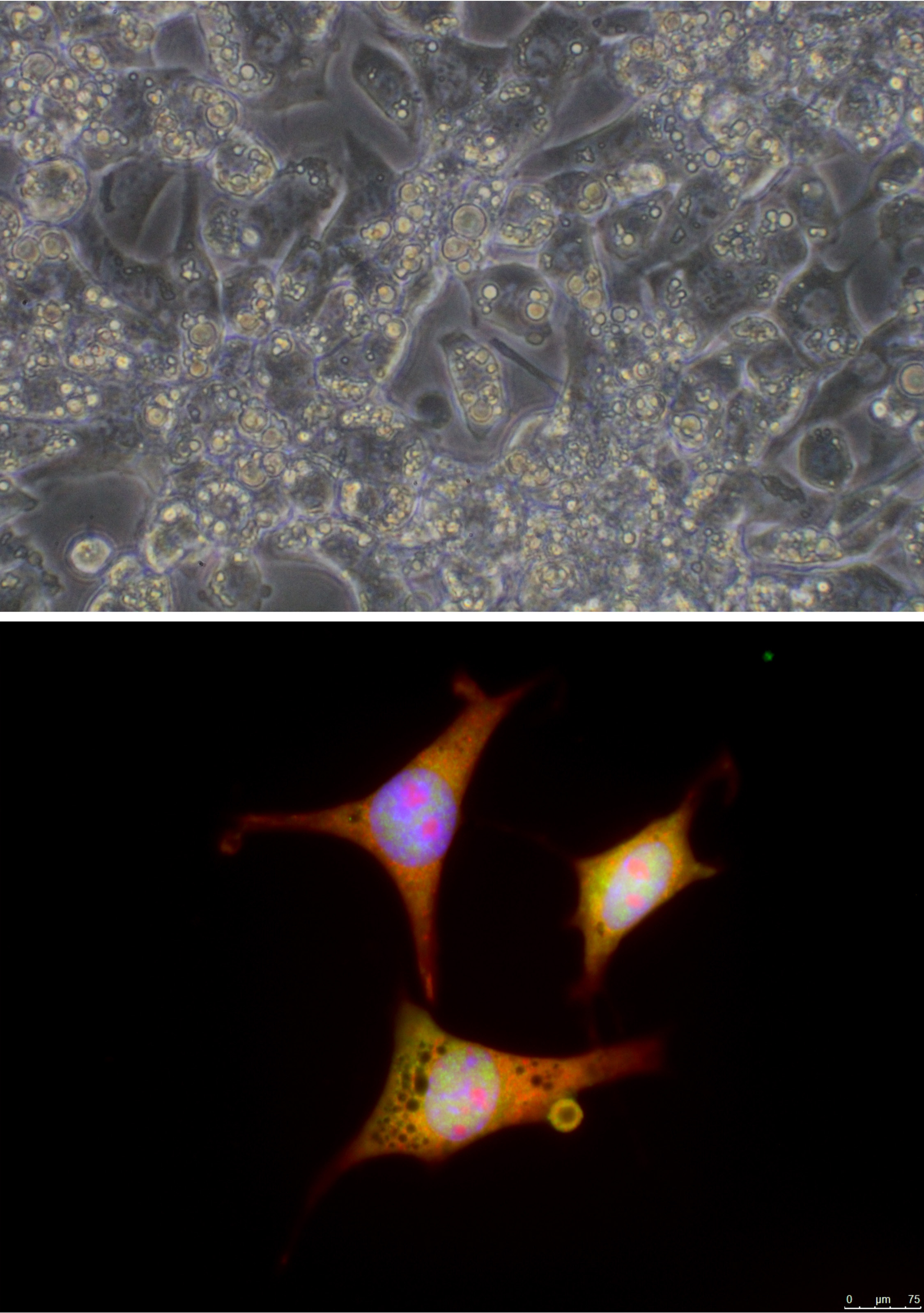 Adipocytes (fat cells) are grown in culture, where intracellular lipid deposits are seen as “bubbles”. Key metabolites (AcetylCoA) and enzymes (ACLY) are visualized in red and green respectively. Images taken by Zyanya Díaz and Tian Gao, research group Verdeguer.
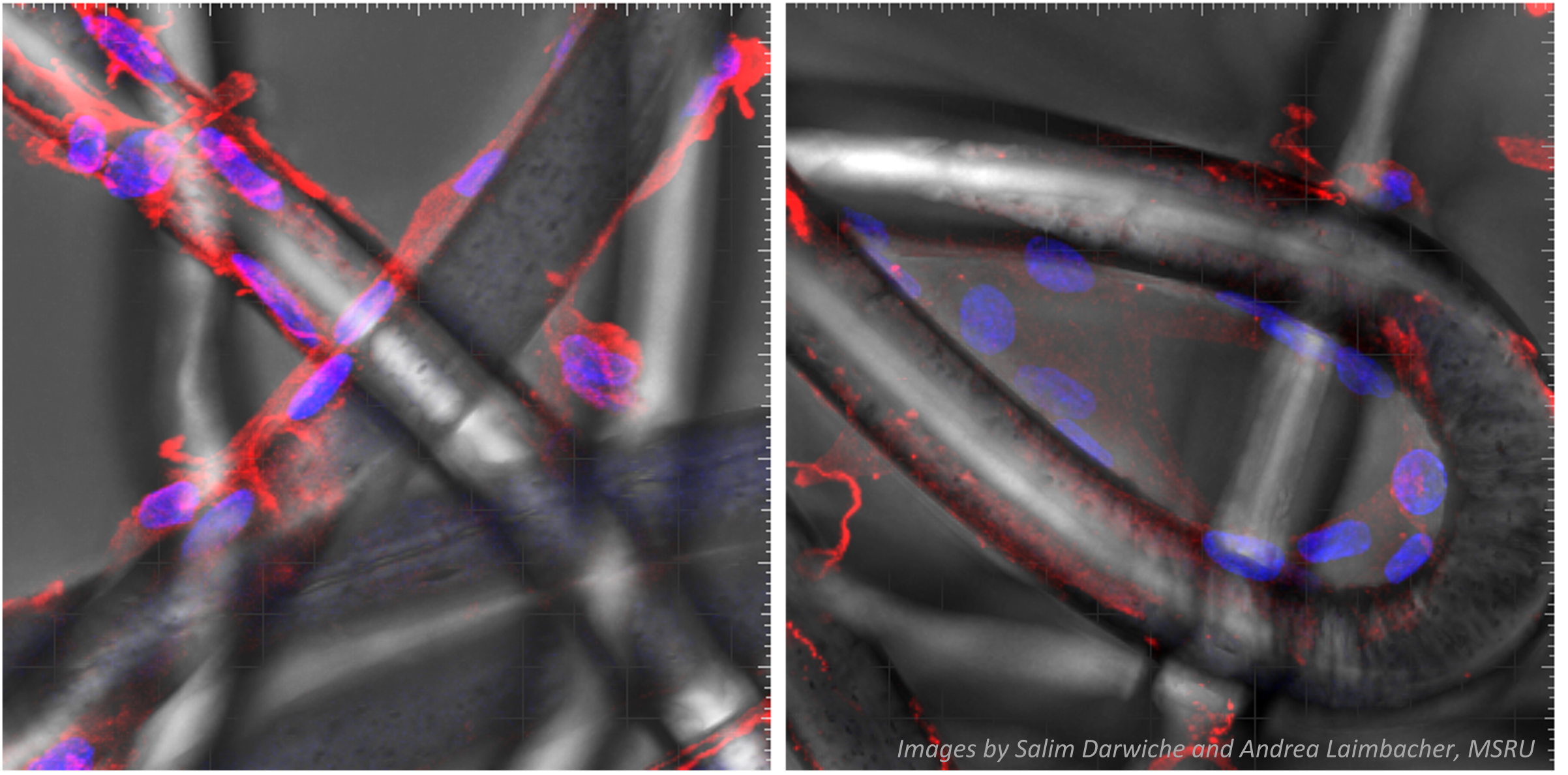 Cells extracted from a sheep’s nucleus pulposus tissue in a cervical intervertebral disc were expanded in culture and seeded on a polyethylene terephthalate scaffold. The fibers are shown above as well as the cell actin cytoskeleton in red and the cell nuclei in blue. The aim of this project is to tissue engineer intervertebral disc components and to implant these constructs for intervertebral disc replacement. Images taken by Salim Darwiche and Andrea Laimbacher, MSRU
Related contributions